Breast Health
You do not have permission to view discussions in this forum.
-
Medicine CoursesBreast HealthResume0% complete0% complete
Introduction
Show More Show LessThis Breast Health introductory course provides foundational theoretical and practical knowledge and skills including how to provide effective counseling regarding breast health. All components of this training are free, including registration, learning, testing, and a certificate of completion. This course is intended for physicians, nurses, and allied health professionals in training and in practice.
-
Students mustStudent must submit this questionnaire to complete it
-
Students mustViewReceive a grade
Show More-
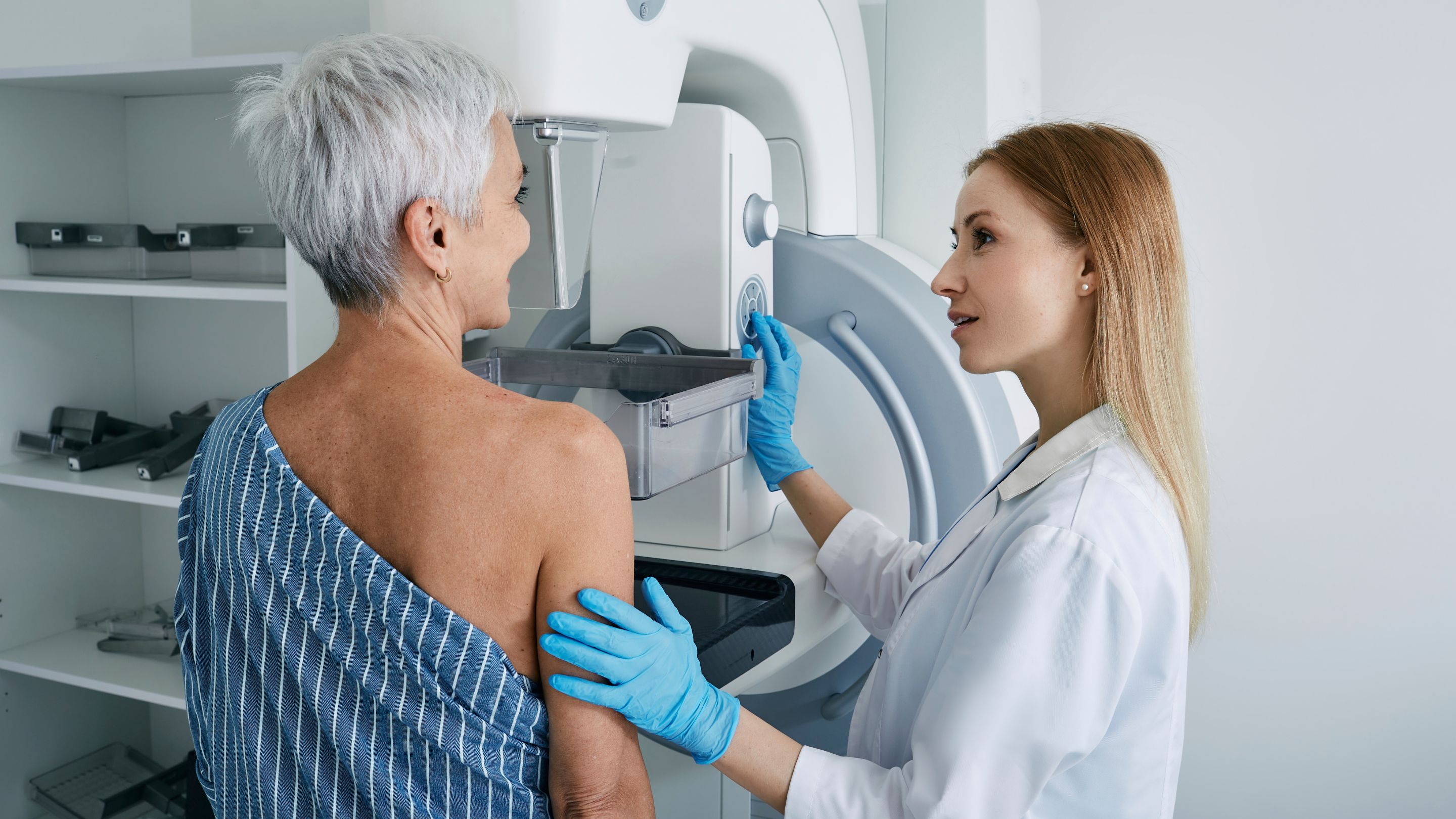

This Breast Health introductory course provides foundational theoretical and practical knowledge and skills including how to provide effective counseling regarding breast health. All components of this training are free, including registration, learning, testing, and a certificate of completion. This course is intended for physicians, nurses, and allied health professionals in training and in practice.
This Breast Health course was developed in partnership with the University of Florida through the affiliation of the course creator, Maryam Sattari, MD, MS, Associate Professor of Medicine, Division of General Internal Medicine. Like all NextGenU courses, this course is competency-based and uses adapted Breast Health competencies outlined in Educational Objectives: Core Curriculum in Obstetrics and Gynecology, 10th Edition (designed by affiliates of the Education Committee of the Council on Resident Education in Obstetrics and Gynecology (CREOG)). It also uses resources from world-class organizations such as Johns Hopkins Medicine, the National Cancer Institute, and the U.S. Preventive Task Force. The course was developed by Maryam Sattari, MD, MS with advice from Erica Frank, MD, MPH, and several faculty members of the University of Florida.
For a publication on this course’s efficacy, see “Building Public Health Capacity through Online Global Learning,” (2018), Open Praxis to see more research related to NextGenU.org’s education model, check out NextGenU.org's publication page. Subscribe to our newsletter to be notified of future updates, new courses, and to be part of our community.
There are 7 modules to complete, which provide:
- A basic introduction to the anatomy and physiology of the breast, benign disorders of the breast;
- Routine screening approaches to selected diseases of the breast;
- Epidemiology and risk assessment of breast cancer;
- Basic mechanisms of genetic inheritance of breast cancer, and diagnosis of breast cancer.
Approximate time required for the required readings for the course is 12 hours at an average reading rate of 144 words/minute; in addition, there are required activities.
The course requires the completion of all peer activities. At the end of each lesson, there is a practice quiz. At the end of the course, after you’ve completed each lesson, quiz, and activity, you’ll have access to a final exam, and a chance to assess the training. Once you’ve passed that last test, you will be able to download a certificate of completion from NextGenU.org and our course’s co-sponsoring organizations (listed above). We keep all of your personal information confidential, never sell any of your information, and only use anonymized data for research purposes, and we are also happy to report your testing information and share your work with anyone (your school, employer, etc.) at your request. We hope that you will find this a rewarding learning experience, and we count on your assessment and feedback to help us improve this training for future students.
Engaging with this Course:
You may browse this course for free to learn for your personal enrichment; there are no requirements.
To obtain a certificate
- Show in the registration fields that you have the appropriate prerequisites to be certified. This course requires a college-level degree.
- Complete all the reading requirements.
- Complete all quizzes.
- Complete 5 peer activity and associated certification quizzes.
- Successfully complete the final exam with a minimum of 70% and a maximum of 3 attempts.
- Complete the self and course evaluation forms.
To obtain credit
- Complete all requirements listed above for the certificate.
- Your learning institution or workplace should approve the partner-university-sponsored NextGenU.org course for educational credit, as they would for their learner taking a course anywhere.
- NextGenU.org is happy to provide your institution with:
- a link to and description of the course training, so they can see all its components, including the cosponsoring universities and other professional organization cosponsors;
- your grade on the final exam;
- your work products (e.g. peer and mentored activities), and any other required or optional shared materials that you produce and authorize to share with them;
- your evaluations -- self-, peer-, and [for mentored courses] mentor assessments;
- a copy of your certificate of completion, with the co-sponsoring universities and other organizations listed.
To obtain a degree co-sponsored with NextGenU.org, registrants must be enrolled in a degree program as a student of a NextGenU.org institutional partner. If you think that your institution might be interested in offering a degree with NextGenU.org contact us.
We hope that you will find this a rewarding learning experience, and we count on your assessment and feedback to help us improve this training for future students.
Next Steps
- Complete the registration form.
- Begin the course with Module 1: Anatomy of the Breast
-
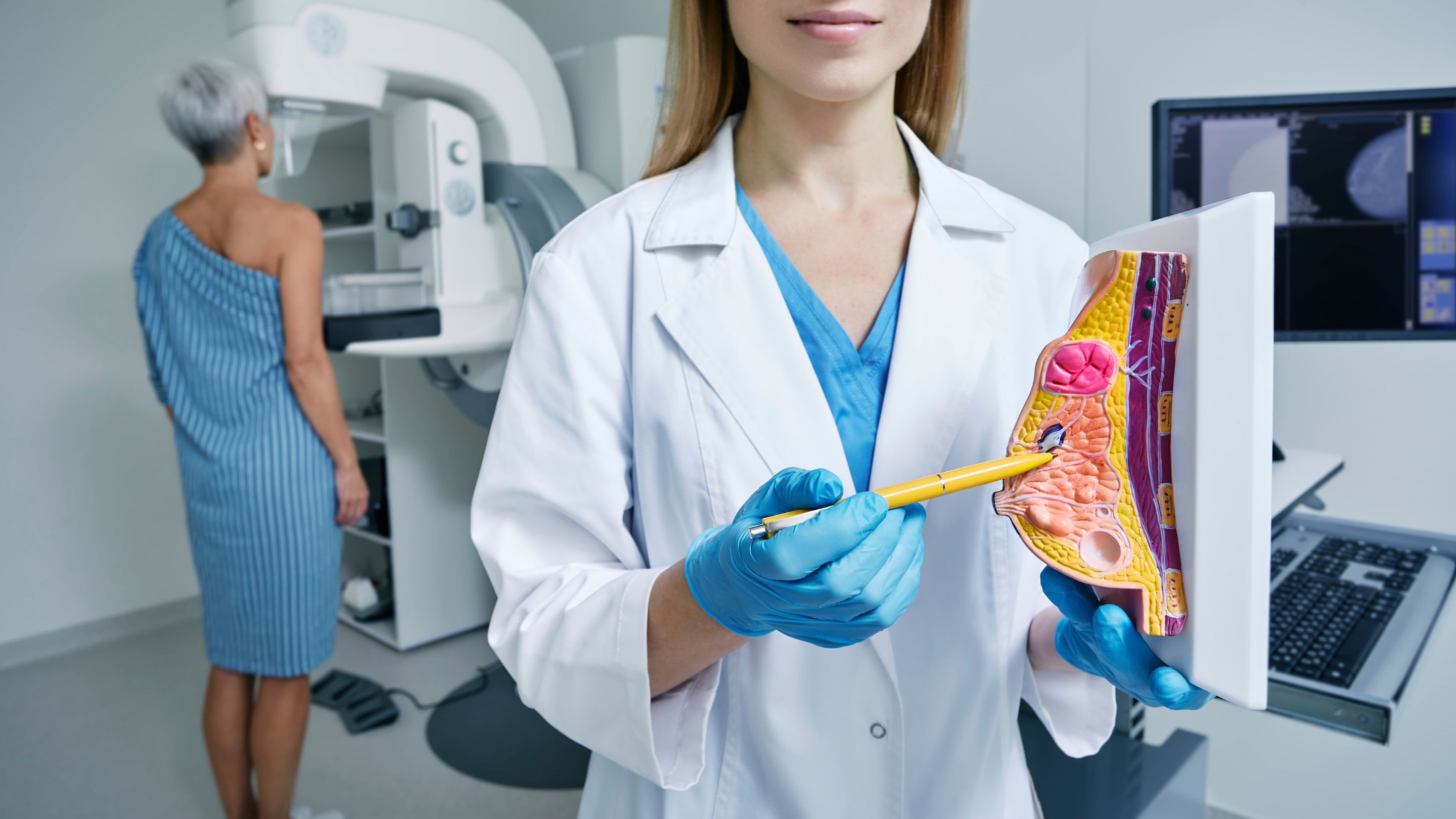 Competency covered in this module:
 - Demonstrate comprehensive knowledge of breast anatomical structures and their clinical significance.
Approximate time required for the readings for this module (at 144 words/minute): 5 hours and 19minutes.-
Students mustMark as done
This module will act as a foundation for the rest of the course, providing you with an understanding of the gross and histologic anatomy of the breast.
-
Students mustMark as done
Required Learning Resources
-
Students mustMark as done
Please review the learning resource summarizing the key concepts and information for Module 1. This resource provides a comprehensive overview, highlighting the most important points and insights to help you grasp the foundational elements of the module.
If you would like more detailed information, please refer to the supplementary links provided below.
263.3 KB -
Students mustMark as done
Supplementary Learning Resources (Not Required)
-
Students mustMark as done
In this document, we summarized key topics of the module to help you study. Please note that these notes do not cover all of the material in its entirety; you are still encouraged to read and study all of the required learning resources.
243.7 KB -
Students mustView
Here are specialized flashcards designed to reinforce and test your knowledge throughout this course.
-
Students mustViewComplete the activity
-
Student Learning Outcomes:
Upon completion of this lesson, you will be able to:- Accurately label all external breast structures on a diagram.
- Describe the relationship between breast tissue layers.
- Explain the lymphatic drainage of the breast
-
Required Learning Resources
-
Students mustView
Read the entire article to learn about the female breast. (10 minutes)
Johns Hopkins Medicine - Pathology
-
Students mustMark as done
Read the entire article to learn about the female breast. (10 minutes)
Health Library Cleveland Clinic - 2023
-
Students mustMark as done
Read the entire article to learn about the lymphatic drainage of the breast. (10 minutes)
Radiopedia - 2023
-
Students mustMark as done
Read the entire article. (10 minutes)
MDPI - 2021
-
Student Learning Outcomes:
Upon completion of this lesson, you will be able to:- Correctly identify cellular structures on histologic slides.
- Draw and label a complete breast ductal system.
- Explain the significance of the TDLU in breast pathology.
-
Required Learning Resources
-
Students mustMark as done
Click on the corresponding links to access plates 13.253-13.256. Examine the plates and read the accompanying text to learn about the microscopic anatomy of the mammary gland. (13 minutes)
Michael P. D'Alessandro, M.D., and Ronald A. Bergman, Ph.D curate Anatomy Atlases - 2015
-
Students mustMark as done
Read, Draw and label a complete breast ductal system. (5 minutes)
Barbara Luz Oliveira - 2018
-
Students mustMark as done
Read the entire article. (15 minutes)
1996 - 2019 Humpath.com - Human Pathology - 2016
-
Students mustViewStart discussions: 1
SLOs:
Accurately label all external breast structures on a diagram
Describe the relationship between breast tissue layers
Explain the lymphatic drainage of the breast
Correctly identify cellular structures on histologic slides
Draw and label a complete breast ductal system
Explain the significance of the TDLU in breast pathology
General Instructions:
In this activity, you will demonstrate your understanding of breast anatomy through critical analysis and application of anatomical knowledge to clinical scenarios.
Step 1: Select and Reflect
First, review the slide sets on "Gross Anatomy of the Breast" and "Histologic Anatomy of the Breast." These will provide you with essential information about external breast structures, tissue layers, lymphatic drainage, and cellular components.
Then read the article "Clinical Implications of Breast Anatomy and Development" by Hassiotou & Geddes. This will help you understand the relationship between anatomical structures and their clinical significance.
You may also wish to review additional resources on breast anatomy and histology for a deeper understanding of the subject.
Step 2: Critical Analysis and Response
Based on your readings and research, compose a 600-800 word response, including the use of diagrams, addressing the following:
Analyze the clinical significance of lymphatic drainage patterns of the breast in relation to metastatic spread of breast cancer. Discuss specific anatomical pathways and their implications for staging and surgical management. Explain how detailed knowledge of breast anatomy influences clinical decision-making in surgical approaches and interpretation of imaging studies.
Compare and contrast the anatomical and histological features of the breast across different life stages (adolescence, reproductive years, pregnancy/lactation, and post-menopause). Discuss how these changes impact clinical assessment and interpretation of imaging studies.
Critically evaluate the role of the Terminal Ductal Lobular Unit (TDLU) in breast pathology, including its significance in the development of both benign and malignant conditions.Illustrate your response with a diagram of the complete breast ductal system with labels.
Diagram Requirement: Create one comprehensive diagram that illustrates and labels:
External anatomical breast structures (nipple, areola, Cooper's ligaments, pectoralis major muscle, etc.)
Complete breast ductal system including major ducts, lactiferous ducts, and Terminal Ductal Lobular Units (TDLU)
Histological features of one selected breast life stage showing cellular components such as epithelial cells, myoepithelial cells, and stromal tissue
Diagram Instructions: Create your diagram using digital tools such as Google Drawings, Canva, Lucidchart, or similar online drawing platforms. You may also use tablet drawing apps or scan hand-drawn diagrams. Ensure all structures are clearly labeled with text boxes and connecting lines. Save your diagram as an image file (PNG or JPG) and insert it directly into your Google Document response. Include a brief legend below your diagram if using symbols or color coding.
Step 3: Share
To share your work, click on the "Add a new discussion topic" button under this post and copy/paste your complete Google Document content (including the embedded diagram) into the "Message" box. Make sure to reference others' intellectual property when necessary. All references should follow 7th Edition APA formatting. For further instructions, see the resource on the Himmelfarb Health Sciences Library: APA citation resource (N.B.: references are excluded from word counts).
-
Quiz: Module 1
To access the quiz, click on the name of the quiz provided above. On the following screen, click the attempt quiz button to view the case studies and respond to the questions.
TO PASS THIS QUIZ YOU MUST OBTAIN A SCORE OF 80%.
Not available unless: The activity Pre-Test is marked complete ...Not available unless:- The activity Pre-Test is marked complete
- The activity Course Registration is marked complete
-
 Competency covered in this module:
 - Apply knowledge of breast physiology to understand normal development and function throughout the lifespan.
Approximate time required for the readings for this module (at 144 words/minute): 7 hours and 25 minutes.-
Students mustMark as done
This module will act as foundation for the rest of the course, providing you with an understanding of the physiology of the breast.
-
Students mustMark as done
Required Learning Resources
-
Students mustMark as done
Please review the learning resource summarizing the key concepts and information for Module 1. This resource provides a comprehensive overview, highlighting the most important points and insights to help you grasp the foundational elements of the module.
If you would like more detailed information, please refer to the supplementary links provided below.
330.8 KB -
Students mustMark as done
Supplementary Learning Resources (Not Required)
-
Students mustMark as done
In this document, we summarized key topics of the module to help you study. Please note that these notes do not cover all of the material in its entirety; you are still encouraged to read and study all of the required learning resources.
271.1 KB -
Students mustView
Here are specialized flashcards designed to reinforce and test your knowledge throughout this course.
-
Students mustMark as done
- Apply knowledge of breast physiology to understand normal development and function throughout the lifespan.
-
Student Learning Outcomes:
Upon completion of this lesson, you will be able to:- Outline the key stages of embryonic breast development.
- Explain hormonal influences on pubertal breast development.
- Describe breast tissue changes from adulthood through senescence.
-
Required Learning Resources
-
Students mustView
Read the entire article. (30 minutes)
Published by Cold Spring Harbor Laboratory Press, Pamela Cowin and John Wysolmerski - 2025
-
Students mustView
Read the entire article. (20 minutes)
Dr. Maria Gomez
-
Students mustView
Read the entire article. (30 minutes)
Acta Biochim Biophys Sin (Shanghai) - 2023
- Outline the key stages of embryonic breast development.
-
Student Learning Outcomes:
Upon completion of this lesson, you will be able to:- Describe and explain how hormonal fluctuations at different life stages influence structural and functional changes in breast tissue.
- Explain the preparation of breast tissue for lactation.
- Develop management plans for common lactation-related conditions.
-
Required Learning Resources
-
Students mustView
Read the entire article. (15 minutes)
Pol J Radiol - 2024
-
Students mustView
Read the entire article. (15 minutes)
Fastest Obstetric, Gynecology, and Pediatric Insight Engine - 2025
-
Students mustView
Read the entire article. (15 minutes)
Joshua Napilay - 2024
-
Students mustViewStart discussions: 1
SLOs:
Outline the key stages of embryonic breast development
Explain hormonal influences on pubertal breast development
Describe breast tissue changes from adulthood through senescence
Describe and explain how hormonal fluctuations at different life stages influence structural and functional changes in breast tissue
Explain the preparation of breast tissue for lactation
Develop management plans for common lactation-related conditions
General Instructions:
In this activity, you will demonstrate your understanding of breast physiology through the development of an educational resource and critical peer review.
Step 1: Research and Preparation
First, review the slide sets on "Breast Development" and "Reproductive Cycle Changes." These will provide you with foundational knowledge about embryonic development, pubertal changes, and reproductive cycle influences on breast tissue.
Then read the articles "Hormonal Regulation of Mammary Development and Milk Production" and "Breast Changes Throughout the Lifespan: A Comprehensive Review."
You may also wish to review additional resources on breast physiology and lactation management for a more comprehensive understanding.
Step 2: Create an Educational Resource
Develop an educational resource (800-1000 words) designed for healthcare professionals that addresses the following:
Create a comprehensive timeline of breast development and physiological changes from embryonic stage through senescence, highlighting key hormonal fluctuations, and specific structural and functional tissue changes (e.g., ductal proliferation, lobular development, stromal changes).
Explain the physiological preparation of breast tissue for lactation, including cellular changes and hormonal regulation.
Design a decision-making algorithm for the assessment and management of one common lactation-related condition (e.g., engorgement, mastitis, insufficient milk production), including evidence-based interventions.
Critically discuss how knowledge of normal breast physiology can inform clinical assessment and management of breast-related complaints across different life stages.
Your educational resource should be evidence-based, clinically relevant, and organized in a manner that facilitates learning and application in clinical practice. Ensure it is visually appealing. You can choose to create a PowerPoint, poster or blog post.
Step 3: Share
To share your work, click on the "Add a new discussion topic" button under this post and copy/paste your complete Google Document content into the "Message" box. Make sure to reference others' intellectual property when necessary. All references should follow 7th Edition APA formatting. For further instructions, see the resource on the Himmelfarb Health Sciences Library: APA citation resource (N.B.: references are excluded from word counts).
-
Quiz: Module 2
To access the quiz, click on the name of the quiz provided above. On the following screen, click the attempt quiz button to view the case studies and respond to the questions.
TO PASS THIS QUIZ YOU MUST OBTAIN A SCORE OF 80%.
Not available unless: The activity Quiz: Module 1 is marked complete
- Describe and explain how hormonal fluctuations at different life stages influence structural and functional changes in breast tissue.
-
 Competency covered in this module:
 - Evaluate, diagnose, and manage common benign breast disorders using evidence-based approaches.
Approximate time required for the readings for this module (at 144 words/minute): 19 hours and 25 minutes.-
Students mustMark as done
After reading the resources and participating in the activities in this module, you will become familiar with the presentation of benign breast disorders, their work-up, treatment, and management.
-
Required Learning Resources
-
Students mustMark as done
Please review the learning resource summarizing the key concepts and information for Module 1. This resource provides a comprehensive overview, highlighting the most important points and insights to help you grasp the foundational elements of the module.
If you would like more detailed information, please refer to the supplementary links provided below.
420.5 KB -
Students mustMark as done
Supplementary Learning Resources (Not Required)
-
Students mustMark as done
In this document, we summarized key topics of the module to help you study. Please note that these notes do not cover all of the material in its entirety; you are still encouraged to read and study all of the required learning resources.
258.5 KB -
Students mustView
Here are specialized flashcards designed to reinforce and test your knowledge throughout this course.
-
Students mustMark as done
-
Student Learning Outcomes:
Upon completion of this lesson, you will be able to:- Differentiate between physiologic and pathologic nipple discharge and galactorrhea.
- List red flag features of nipple discharge that suggest malignancy.
- Describe a structured diagnostic approach to evaluating patients with nipple discharge and galactorrhea.
Click here to start this lesson-
Required Learning Resources
-
-
Students mustView
Read the entire article. (15 minutes)
NIH - National Library of Medicine - 2023
-
Students mustView
Read the entire article to learn about nipple discharge. (30 minutes)
Lydia Choi, MD, Karmanos Cancer Center - 2025
-
Students mustView
Read the entire article. (10 minutes)
NLM provides: Veda Parthasarathy, Usharani Rathnam - 2012
-
Students mustView
Read the entire article. (30 minutes)
American Journal of Roentgenology - 2020
-
Students mustView
Read the entire article. (30 minutes)
NLM provides: Marco Moschetta , Vincenzo De Ruvo - 2020
-
Student Learning Outcomes:
Upon completion of this lesson, you will be able to:- Create a management plan for cyclic mastalgia.
- Diagnose lactational vs. non-lactational mastitis.
- Implement appropriate antibiotic and supportive therapies for mastitis.
Click here to start this lesson-
Required Learning Resources
-
Students mustView
Read the entire article. (10 minutes)
Health Central By Alyssa Sparacino Medical Reviewer Andrea Eisenberg, M.D. - 2024
-
Students mustView
Read the entire article. (30 minutes)
C. Gopalakrishnan Nair Hiran - 2015
-
Students mustView
Read the entire article. (15 minutes)
BiologyInsights Team - 2025
-
Students mustView
Read the entire article. (45 minutes)
NLM provides - 2022
-
Students mustMark as done
Supplementary Learning Resources (Not Required)
-
Read the article to learn more about breast pain. (3 minutes)
Mary Ann Kosir, MD - 2016
-
Read the entire article. (3 minutes)
LACTAPP - 2022
-
Student Learning Outcomes:
Upon completion of this lesson, you will be able to:- Differentiate fibroadenoma from other benign masses.
- Evaluate and manage breast asymmetry.
- Demonstrate proper technique for clinical breast examination with appropriate documentation.
Click here to start this lesson-
Required Learning Resources
-
Students mustView
Read the entire article. (20 minutes)
Acibadem Health Point - 2025
-
Students mustView
Read the entire article. (30 minutes)
NLM provides: - 2023
-
Students mustView
Read the entire article. (35 minutes)
Journal of Plastic, Reconstructive & Aesthetic Surgery - 2023
-
Students mustView
Read the entire article. (20 minutes)
Stanford University - 2025
-
Students mustView
Read the entire article. (15 minutes)
NLM provides - 2023
-
Students mustViewStart discussions: 1
SLOs:
Differentiate between physiologic and pathologic nipple discharge and galactorrhea
List red flag features of nipple discharge that suggest malignancy
Describe a structured diagnostic approach to evaluating patients with nipple discharge and galactorrhea
Create a management plan for cyclic mastalgia
Diagnose lactational vs. non-lactational mastitis
Implement appropriate antibiotic and supportive therapies for mastitis
Differentiate fibroadenoma from other benign masses
Evaluate and manage breast asymmetry
Demonstrate proper technique for clinical breast examination with appropriate documentation
General Instructions:
In this activity, you will demonstrate your understanding of benign breast disorders through case analysis and development of evidence-based management plans.
Step 1: Select and Reflect
First, review the slide sets on "Nipple Discharge and Galactorrhea," "Breast Pain and Infections," and "Breast Masses and Structural Abnormalities." These will provide you with foundational knowledge about various benign breast conditions.
Then read the articles "Clinical Management of Benign Breast Disorders" and "Evidence-Based Approaches to Benign Breast Complaints."
You may also wish to review current clinical practice guidelines for the management of benign breast disorders from organizations such as the American College of Obstetricians and Gynecologists (ACOG) or the National Comprehensive Cancer Network (NCCN).
Step 2: Case Analysis and Management Plan
Based on your readings and research, analyze the following case and develop a comprehensive management plan (800-1000 words):
Case Scenario A:
A 32-year-old woman presents with a 3-month history of bilateral breast pain that worsens in the week before her menstrual period. She also reports intermittent clear nipple discharge from her right breast that occurs spontaneously. She recently noticed a mobile, firm, well-circumscribed mass in her left breast that is mildly tender to palpation.She also mentions she has noticed her breasts appear unequal in size over the past year. She has no personal or family history of breast cancer. She has had two pregnancies resulting in two live births and breastfed both children for approximately 6 months each. Her last menstrual period was 2 weeks ago, and she reports regular 28-day cycles.Case Scenario B:
A 28-year-old woman who is six weeks postpartum and currently breastfeeding presents with unilateral breast swelling, localized erythema and warmth, increased pain, and a fever of 38.5°C (101.3°F). She reports reduced drainage from the affected breast over the past 24 hours.She is exclusively breastfeeding and reports the baby has been feeding well. Upon examination, the right breast shows erythema in the upper outer quadrant with a tender, indurated area.
Your response should include:
Provide a differential diagnosis for each of the patient’s main concerns.
For Case A, you must differentiate physiologic from pathologic causes of the patient’s nipple discharge and differentiate fibroadenoma from other benign breast masses when addressing the palpable lesion.
For Case B, identify the differential diagnosis for lactational versus non-lactational mastitis and explain potential causes of breast asymmetry. You must justify each diagnosis with reference to the case details and relevant clinical features.
Describe a structured diagnostic workup for one of these 2 patients. This should include the additional history you would obtain, the components of the physical examination you would perform, and the imaging or laboratory studies you would pursue. As part of your physical examination description, you must demonstrate proper clinical breast examination documentation by describing the findings using clock-face notation, breast quadrants, and the measured distance of findings from the nipple, along with the characteristics of any palpable abnormalities (size, shape, consistency, mobility, skin changes).
For your chosen patient, develop an evidence-based management plan for the most likely diagnoses including pharmacologic and non-pharmacologic interventions. You should reference current clinical guidelines where appropriate. If choosing Case B, your management plan must include specific antibiotic selection, dosing, duration, and supportive therapies for mastitis treatment, explaining your rationale for the chosen approach.
For your chosen patient, discuss the red flag features that would alter your management approach. Identify findings that would raise concern for malignancy or complicated infection and explain how these findings would change your diagnostic or therapeutic plan.
For your chosen patient, draft 1 patient-centered education point that you would address. Your discussion should include guidance on symptom expectations, self-monitoring, breastfeeding recommendations when relevant, supportive strategies, and instructions for when to seek urgent follow-up care.
Step 3: Share
To share your work, click on the "Add a new discussion topic" button under this post and paste your work into the "Message" box. Make sure to reference others' intellectual property when necessary. All references should follow 7th Edition APA formatting. For further instructions, see the resource on the Himmelfarb Health Sciences Library: APA citation resource (N.B.: references are excluded from word counts).
-
Quiz: Module 3
To access the quiz, click on the name of the quiz provided above. On the following screen, click the attempt quiz button to view the case studies and respond to the questions.
TO PASS THIS QUIZ YOU MUST OBTAIN A SCORE OF 80%.
Not available unless: The activity Quiz: Module 2 is marked complete
-
 Competency
covered in this module:
Competency
covered in this module: - Implement and interpret appropriate screening protocols for breast disease detection and management.
Approximate time required for the readings for this module (at 144 words/minute): 5 hours and 34 minutes.-
Students mustMark as done
This module will provide an overview of the screening and management of selected diseases of the breast such as Mastitis, Galactorrhea, Mastodynia, and Breast Masses.
-
Required Learning Resources
-
Students mustMark as done
Please review the learning resource summarizing the key concepts and information for Module 1. This resource provides a comprehensive overview, highlighting the most important points and insights to help you grasp the foundational elements of the module.
If you would like more detailed information, please refer to the supplementary links provided below.
360.1 KB -
Students mustMark as done
Supplementary Learning Resources (Not Required)
-
Students mustMark as done
In this document, we summarized key topics of the module to help you study. Please note that these notes do not cover all of the material in its entirety; you are still encouraged to read and study all of the required learning resources.
293.1 KB -
Students mustView
Here are specialized flashcards designed to reinforce and test your knowledge throughout this course.
-
Students mustMark as done
-
Student Learning Outcomes:
Upon completion of this lesson, you will be able to:- Compare and contrast mammography, ultrasound, and MRI.
- Apply appropriate screening intervals based on the new recommendations starting at age 40.
- Discuss false positive/negative rates of screening methods.
Click here to start this lesson-
Required Learning Resources
-
Students mustView
Read the entire article. (10 minutes)
Cedars-Sinai Blog - 2021
-
Students mustView
Read the entire article. (15 minutes)
JAMA Netw Open - 2024
-
Students mustView
Read the entire article. (10 minutes)
American Cancer Society - 2022
-
Student Learning Outcomes:
Upon completion of this lesson, you will be able to:- Create a diagnostic plan for a newly discovered breast mass.
- Implement evidence-based treatments for breast pain.
- Identify and manage endocrine causes of galactorrhea.
Click here to start this lesson-
Required Learning Resources
-
Students mustView
Read the entire article. (13 minutes)
Hospital Handbook - 2025
-
Students mustView
Read the entire article. (15 minutes)
Mayo Clin Proc. - 2020
-
Students mustView
Read the entire article. (15 minutes)
John D. Carmichael, MD, Keck School of Medicine of the University of Southern California - 2023
-
Students mustView
Read the entire article. (10 minutes)
NLM provides: Kashif Aziz, Amir Shahbaz - 2018
-
Students mustViewStart discussions: 1
SLOs:
Compare and contrast mammography, ultrasound, and MRI
Apply appropriate screening intervals based on the new recommendations starting at age 40
Discuss false positive/negative rates of screening methods
Create a diagnostic plan for a newly discovered breast mass
Implement evidence-based treatments for breast pain
Identify and manage endocrine causes of galactorrhea
General Instructions:
In this activity, you will demonstrate your understanding of breast cancer screening principles and management of benign breast findings through critical analysis of current guidelines and application to clinical scenarios.
Step 1: Select and Reflect
First, review the slide sets on "Breast Cancer Screening Principles" and "Management of Benign Breast Findings." These will provide you with foundational knowledge about screening modalities and approaches to common breast complaints.
Then read the 2024 USPSTF breast cancer screening guidelines and the accompanying evidence review.
Also review the article "Comparative Effectiveness of Breast Cancer Screening Modalities" and "Management of Common Breast Findings: An Evidence-Based Approach."
You may also wish to review additional resources on breast cancer screening and diagnostic approaches for a more comprehensive understanding.
Step 2: Critical Analysis and Response
Based on your readings and research, compose a 700-900 word response addressing the following:
Critically analyze the 2024 USPSTF breast cancer screening guidelines, including the rationale for starting routine screening at age 40. Compare these recommendations with those of at least two other professional organizations (e.g., American Cancer Society, American College of Radiology, etc.). Explain differences in recommended screening intervals and identify the evidence these organizations prioritize when forming their guidelines.
Compare and contrast mammography, ultrasound, and MRI in both screening and diagnostic contexts. In your analysis, discuss sensitivity, specificity, false-positive and false-negative rates, cost-effectiveness, and clinical scenarios in which one modality is preferred over the others.
Case study 1: You are evaluating a 45-year-old woman with a newly discovered 1.5 cm palpable breast mass. Outline a comprehensive step-by-step diagnostic plan that reflects current guidelines. Include specific imaging selection and sequencing, clear indications for biopsy, and any relevant risk-based considerations. Your plan should demonstrate a systematic approach from initial evaluation through definitive diagnosis.
Case study 2: A separate patient presents with noncyclic breast pain and scant milky nipple discharge. Identify endocrine causes of galactorrhea (including prolactinomas, hypothyroidism, and medication-induced causes) and describe your diagnostic and management approach, including appropriate laboratory evaluation (prolactin levels, TSH, comprehensive medication review) and evidence-based pharmacologic and non-pharmacologic treatments for breast pain.
Briefly discuss the ethical considerations associated with breast cancer screening programs, including overdiagnosis, overtreatment, and psychological distress associated with false positives. Propose 2 evidence-based strategies to mitigate these harms while preserving early-detection benefits.
Step 3: Share
To share your work, click on the "Add a new discussion topic" button under this post and paste your work into the "Message" box. Make sure to reference others' intellectual property when necessary. All references should follow 7th Edition APA formatting. For further instructions, see the resource on the Himmelfarb Health Sciences Library: APA citation resource (N.B.: references are excluded from word counts).
-
Quiz: Module 4
To access the quiz, click on the name of the quiz provided above. On the following screen, click the attempt quiz button to view the case studies and respond to the questions.
TO PASS THIS QUIZ YOU MUST OBTAIN A SCORE OF 80%.
Not available unless: The activity Quiz: Module 3 is marked complete
-
 Competency covered in this module:
 - Apply epidemiological principles to assess breast cancer risk and develop comprehensive risk reduction strategies.
Approximate time required for the readings for this module (at 144 words/minute): 20 hours and 25 minutes.-
Students mustMark as done
This module will provide you with an overview of the epidemiology of breast cancer as well as the evaluation and effective counseling of patients with a risk for breast cancer.
-
Required Learning Resources
-
Students mustMark as done
Please review the learning resource summarizing the key concepts and information for Module 1. This resource provides a comprehensive overview, highlighting the most important points and insights to help you grasp the foundational elements of the module.
If you would like more detailed information, please refer to the supplementary links provided below.
340.3 KB -
Students mustMark as done
Supplementary Learning Resources (Not Required)
-
Students mustMark as done
In this document, we summarized key topics of the module to help you study. Please note that these notes do not cover all of the material in its entirety; you are still encouraged to read and study all of the required learning resources.
273.9 KB -
Students mustView
Here are specialized flashcards designed to reinforce and test your knowledge throughout this course.
-
Students mustMark as done
-
Click here to start this lesson
Student Learning Outcomes:
Upon completion of this lesson, you will be able to:- Counsel patients on modifiable risk factors.
- Develop tailored screening plans for patients with increased risk based on race/ethnicity.
- Demonstrate proficiency with risk assessment tools (Gail, Tyrer-Cuzick, etc.)
-
Required Learning Resources
-
Students mustView
Read the sections titled "Who's at risk?" and "Overview" to learn about the factors associated with increased and decreased risk of breast cancer, as well as the interventions that reduce the risk of breast cancer. (14 minutes)
National Cancer Institute - 2025
-
Students mustView
Read the entire article. (45 minutes)
JAMA Network Open - 2025
-
Students mustView
Read the entire article to learn about the importance of a personal history of breast cancer. (20 minutes)
Schacht DV, Yamaguchi K, Lai J, Kulkarni K, Sennett CA, Abe H. -2014
-
Students mustView
Read the section titled "Inherited Risk" and click on the 5 links in this section to read about breast cancer risk assessment tools. (15 minutes)
National Cancer Institute - 2025
-
Click here to start this lesson
Student Learning Outcomes:
Upon completion of this lesson, you will be able to:- Develop personalized risk reduction plans.
- Identify appropriate candidates for SERMs and aromatase inhibitors.
- Counsel patients on the risk/benefit of prophylactic surgery.
-
Required Learning Resources
-
Students mustView
Read the entire article. (40 minutes)
The Journal of Family Practice - 2012
-
Students mustView
Read the entire article. (35 minutes)
NIH - National Library of Medicine - 2021
-
Students mustView
Read the entire article. (35 minutes)
Cochrane Breast Cancer Group - 2019
-
Students mustView
Read the entire article. (15 minutes)
National Cancer Institute - 2025
-
Click here to start this lesson
Student Learning Outcomes:
Upon completion of this lesson, you will be able to:- Determine appropriate candidates for breast MRI and supplemental screening.
- Create individualized screening plans for high-risk patients.
- Refer appropriate patients for genetic counseling based on updated criteria.
-
Required Learning Resources
-
Students mustView
Read pages 96-103 and review Table 2 for information related to breast cancer. (60 minutes)
CA: a Cancer Journal for Clinicians - 2016
-
Students mustView
Read the web page to understand the current recommendations for breast cancer screening as well as the controversies and differences in recommendations by different expert societies and guidelines. (36 minutes)
U.S. Preventive Services Task Force - 2016
-
Students mustView
Read the web page to understand the breast cancer screening for High-Risk Women. (15 minutes)
American College of Radiology- 2023
-
Students mustView
Read the entire article and see the YouTube video. (15 minutes)
CDC - 2023
-
Students mustView
Read the entire article. (40 minutes)
USPSTF Recommendations App - Prevention Task Force - 2019
-
Students mustViewStart discussions: 1
SLOs:
Counsel patients on modifiable risk factors
Develop tailored screening plans for patients from populations experiencing disproportionate disease burden due to social determinants of health and structural inequities.
Demonstrate proficiency with risk assessment tools (Gail, Tyrer-Cuzick, etc.)
Develop personalized risk reduction plans
Identify appropriate candidates for SERMs and aromatase inhibitors
Counsel patients on risk/benefit of prophylactic surgery
Determine appropriate candidates for breast MRI and supplemental screening
Create individualized screening plans for high-risk patients
Refer appropriate patients for genetic counseling based on updated criteria
General Instructions:
In this activity, you will demonstrate your understanding of breast cancer risk assessment and risk reduction strategies through the development of personalized risk management plans and peer evaluation.
Step 1: Research and Preparation
First, review the slide sets on "Breast Cancer Risk Factors," "Risk Reduction Strategies," and "Screening in High-Risk Populations." These will provide you with foundational knowledge about risk assessment and management.
Then read the articles "Current Approaches to Breast Cancer Risk Assessment" and "Evidence-Based Risk Reduction Strategies."
Familiarize yourself with at least two breast cancer risk assessment tools (e.g., Gail Model, Tyrer-Cuzick, Breast Cancer Surveillance Consortium Risk Calculator).
Review the current NCCN guidelines for breast cancer risk reduction and screening for high-risk individuals.
Step 2: Case Analysis and Risk Management Plans
Develop risk assessment and management plans (800-1000 words total) for TWO of the following case scenarios:
Case 1: A 42-year-old African American woman with no personal history of breast cancer. Her mother was diagnosed with breast cancer at age 58, and she has one sister who is breast cancer-free. She began menstruating at age 11, had her first child at age 32, and has never used hormone replacement therapy. She has a BMI of 31 and drinks 5-7 alcoholic beverages per week. She has never had a breast biopsy.
Case 2: A 35-year-old Hispanic woman whose maternal grandmother and maternal aunt were diagnosed with breast cancer at ages 45 and 50, respectively. She has no personal history of contraceptives for 15 years. She has a BMI of 24 and exercises regularly. She had a breast biopsy at age 30 that showed atypical ductal hyperplasia.
Case 3: A 55-year-old Caucasian woman who tested positive for a BRCA1 mutation. She has no personal history of breast or ovarian cancer. Her mother and sister both had breast cancer in their 40s. She began menstruating at age 12, had two children before age 30, and went through menopause at age 52. She has a BMI of 27 and is currently taking medication for hypertension.
For each of your 2 selected cases, your management plan should include:
A quantitative risk assessment using at least two validated tools, with justification for tool selection, comparison of outputs, and discussion of limitations and uncertainty in calculated risk.
An analysis of modifiable and non-modifiable risk factors, including explanation of how you would counsel this patient about modifiable risk reduction strategies. Integrate relevant population-level data on breast cancer incidence and outcomes while critically examining how social determinants of health influence risk and care for patients from marginalized communities."
A personalized risk reduction plan that incorporates patient-specific lifestyle modifications, chemoprevention strategies, and consideration of prophylactic surgery when appropriate. For chemoprevention, determine candidacy for SERMs or aromatase inhibitors and justify your recommendation with specific criteria.
A tailored screening plan with justification for the recommended modalities and intervals, including the evidence-based criteria used to determine eligibility for breast MRI or other supplemental screening.
Genetic counseling and testing recommendations based on updated NCCN criteria, including an explanation of which criteria the patient meets and how these influence management decisions.
One patient-centered counseling point, such as discussion of risks and benefits of preventive strategies, addressing patient concerns, clarifying misconceptions, and incorporating patient preferences into shared decision-making.
Step 3: Share
To share your work, click on the "Add a new discussion topic" button under this post and paste your work into the "Message" box. Make sure to reference others' intellectual property when necessary. All references should follow 7th Edition APA formatting. For further instructions, see the resource on the Himmelfarb Health Sciences Library: APA citation resource (N.B.: references are excluded from word counts).
-
Quiz: Module 5
To access the quiz, click on the name of the quiz provided above. On the following screen, click the attempt quiz button to view the case studies and respond to the questions.
TO PASS THIS QUIZ YOU MUST OBTAIN A SCORE OF 80%.
Not available unless: The activity Quiz: Module 4 is marked complete
-
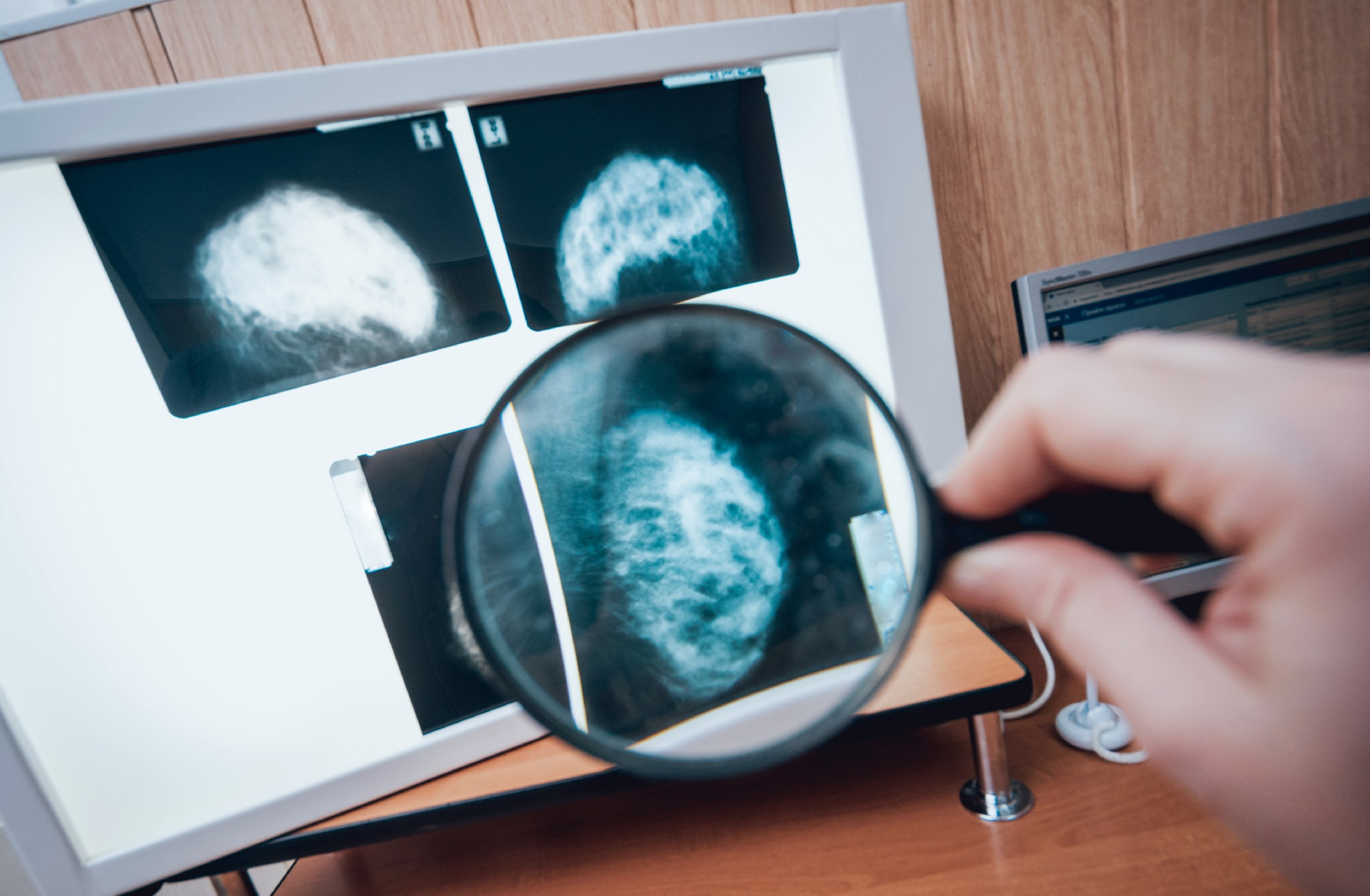 Competency covered in this module:
 - Integrate genetic principles into breast cancer risk assessment and management.
Approximate time required for the readings for this module (at 144 words/minute): 5 hours and 58 minutes.-
Students mustMark as done
In this module, you will learn about common inheritance patterns of breast cancer and indications for screening for certain genes related to breast cancer.
-
Required Learning Resources
-
Students mustMark as done
Please review the learning resource summarizing the key concepts and information for Module 1. This resource provides a comprehensive overview, highlighting the most important points and insights to help you grasp the foundational elements of the module.
If you would like more detailed information, please refer to the supplementary links provided below.
289.8 KB -
Students mustMark as done
Supplementary Learning Resources (Not Required)
-
Students mustMark as done
In this document, we summarized key topics of the module to help you study. Please note that these notes do not cover all of the material in its entirety; you are still encouraged to read and study all of the required learning resources.
263.9 KB -
Students mustView
Here are specialized flashcards designed to reinforce and test your knowledge throughout this course.
-
Students mustMark as done
-
Student Learning Outcomes:
Upon completion of this lesson, you will be able to:- Differentiate between dominant and recessive inheritance.
- Explain variable penetrance in BRCA mutations.
- Recognize clinical features of hereditary breast cancer syndromes, including PALB2, ATM, and CHEK2.
-
Required Learning Resources
-
Students mustMark as done
Read the sections titled "Description", "Frequency", "Causes", and "Inheritance Pattern". This resource provides information about hereditary breast cancer. (9 minutes)
National Institutes of Health, U.S. National Library of Medicine - 2015
-
Students mustMark as done
Read the entire article. (20 minutes)
npj Precision Oncology volume - 2024
-
Students mustMark as done
Supplementary Learning Resources (Not Required)
-
Students mustMark as done
Read the article to learn more about the genetics of breast cancer. (31 min)
S. Shiovitz and L. A. Korde - 2015
-
Student Learning Outcomes:
Upon completion of this lesson, you will be able to:- Select appropriate patients for genetic testing based on updated NCCN criteria.
- Describe different genetic testing methodologies, including VUS interpretation.
- Develop management plans based on genetic findings.
-
Required Learning Resources
-
Students mustMark as done
Read the entire article. (15 minutes)
NLM provides - 2021
-
Students mustMark as done
Read the entire recommendation statement to understand the current indications for screening for BRCA1 and BRCA2. (42 minutes)
U.S. Preventive Services Task Force. - 2019
-
Students mustViewStart discussions: 1
SLOs:
Differentiate between dominant and recessive inheritance
Explain variable penetrance in BRCA mutations
Recognize clinical features of hereditary breast cancer syndromes including PALB2, ATM, and CHEK2
Select appropriate patients for genetic testing based on updated NCCN criteria
Describe different genetic testing methodologies including VUS interpretation
Develop management plans based on genetic findings
General Instructions:
In this activity, you will demonstrate your understanding of genetic principles in breast cancer risk assessment and management through the development of educational materials and peer evaluation.
Step 1: Research and Preparation
First, review the slide sets on "Principles of Cancer Genetics" and "Genetic Testing and Counseling." These will provide you with foundational knowledge about inheritance patterns, gene penetrance, and genetic testing methodologies.
Then read the articles "Hereditary Breast Cancer: Beyond BRCA" and "Clinical Management of Patients with Variants of Uncertain Significance."
Review the current NCCN guidelines for genetic/familial high-risk assessment for breast, ovarian, and pancreatic cancer.
You may also wish to review additional resources on cancer genetics and genetic testing for a more comprehensive understanding.
Step 2: Develop Educational Materials
Develop 2 educational materials (800-1000 words) for healthcare professionals that address the following:
Your responses must demonstrate clinical reasoning and application of genetic principles, not just summary of content.
Create a guide to hereditary breast cancer syndromes that includes the following:
Define and differentiate between Autosomal dominant inheritance and Autosomal recessive inheritance. Explain why BRCA-related breast cancer follows dominant inheritance
Include a sample three-generation pedigree demonstrating autosomal dominant inheritance in a breast cancer family with annotations showing:
Multiple affected individuals across generations
~50% transmission pattern
Unaffected carriers (demonstrating incomplete penetrance)
Explain how inheritance pattern informs genetic counseling (50% risk for first-degree relatives) and cascade testing strategy
Include a comparison table of at least five hereditary breast cancer syndromes (including BRCA1/2, PALB2, ATM, and CHEK2), their inheritance patterns, estimated cancer risks, associated clinical features, and key evidence-based surveillance recommendations
Explain variable penetrance and expressivity with specific examples related to breast cancer genes
A discussion of gene-environment interactions in hereditary breast cancer
Introduce risk-reducing intervention options with their efficacy and limitations and cascade testing recommendations for family members.
Develop a clinical decision algorithm for genetic testing referral and interpretation that includes:
Outline the current NCCN criteria for genetic testing referral with specific attention to updates in the most recent guidelines
Describe the following genetic testing technologies:
Next-generation sequencing (NGS): What it detects (point mutations, small indels), advantages (multi-gene panels, cost-effective), limitations (may miss large deletions/duplications)
Deletion/duplication analysis (MLPA or aCGH): What it detects (large rearrangements), why needed (~5-10% of BRCA mutations are large deletions missed by sequencing alone)
Sanger sequencing: When used (confirmation, known familial variants)
Explain the following test selection approaches
Single-gene testing: When appropriate (known familial variant, specific syndrome suspicion)
Multi-gene panel testing: Advantages (comprehensive, identifies unexpected mutations), disadvantages (higher VUS rate)
Panel composition: Which genes to include based on clinical presentation
A structured framework for interpreting and managing variants of uncertain significance (VUS)
Your educational materials should be evidence-based, clinically relevant, and organized in a manner that facilitates learning and application in clinical practice. Include visual elements (tables, algorithms, or decision trees) to enhance understanding.
Step 3: Share
To share your work, click on the "Add a new discussion topic" button under this post and paste your work into the "Message" box. Make sure to reference others' intellectual property when necessary. All references should follow 7th Edition APA formatting. For further instructions, see the resource on the Himmelfarb Health Sciences Library: APA citation resource (N.B.: references are excluded from word counts).
-
Quiz: Module 6
To access the quiz, click on the name of the quiz provided above. On the following screen, click the attempt quiz button to view the case studies and respond to the questions.
TO PASS THIS QUIZ YOU MUST OBTAIN A SCORE OF 80%.
Not available unless: The activity Quiz: Module 5 is marked complete -
Students mustMark as done
Supplementary Learning Resources (Not Required)
-
Students mustMark as done
Read the entire article "NCCN Issues Updated Guidance on Genetic Testing for Several Cancers". (15 minutes)
HealthCentral LLC - 2024
-
 Competency covered in this module:
 - Utilize appropriate diagnostic methods to evaluate, diagnose, and stage breast cancer.
Approximate time required for the readings for this module (at 144 words/minute): 9 hours and 55 minutes.-
Students mustMark as done
This module will provide you with an overview of diagnostic methods to detect an invasive carcinoma of the breast.
-
Required Learning Resources
-
Students mustMark as done
Please review the learning resource summarizing the key concepts and information for Module 1. This resource provides a comprehensive overview, highlighting the most important points and insights to help you grasp the foundational elements of the module.
If you would like more detailed information, please refer to the supplementary links provided below.
308.8 KB -
Students mustMark as done
Supplementary Learning Resources (Not Required)
-
Students mustMark as done
In this document, we summarized key topics of the module to help you study. Please note that these notes do not cover all of the material in its entirety; you are still encouraged to read and study all of the required learning resources.
251.1 KB -
Students mustView
Here are specialized flashcards designed to reinforce and test your knowledge throughout this course.
-
Students mustMark as done
-
Student Learning Outcomes:
Upon completion of this lesson, you will be able to:- Order appropriate diagnostic imaging studies.
- Recognize suspicious findings on mammography, ultrasound, and MRI.
- Apply BIRADS categories to clinical decision-making and follow-up planning.
-
Required Learning Resources
-
Students mustMark as done
Read the entire article. (35 minutes)
The Lancet - 2025
-
Students mustMark as done
Read the entire article. (45 minutes)
Egyptian Journal of Radiology and Nuclear Medicine - 2020
-
Students mustMark as done
Read the entire article. (15 minutes)
Radiological Society of North America - 2025
-
Student Learning Outcomes:
Upon completion of this lesson, you will be able to:- Select appropriate biopsy technique based on lesion characteristics.
- Interpret comprehensive breast pathology reports.
- Explain the significance of ER, PR, HER2, and Ki-67 status in treatment planning.
-
Required Learning Resources
-
Students mustMark as done
Read the entire article. (35 minutes)
American Cancer Society - 2024
-
Students mustMark as done
Read the entire article. (15 minutes)
American Cancer Society - 2024
-
Students mustMark as done
Read the entire article. (30 minutes)
NIH- Library of Medicine - 2024
-
Students mustViewStart discussions: 1
SLOs:
Order appropriate diagnostic imaging studies
Recognize suspicious findings on mammography, ultrasound, and MRI
Apply BIRADS categories to clinical decision-making and follow-up planning
Select appropriate biopsy technique based on lesion characteristics
Interpret comprehensive breast pathology reports
Explain the significance of ER, PR, HER2, and Ki-67 status in treatment planning
General Instructions:
In this activity, you will demonstrate your understanding of breast cancer diagnosis through analysis of imaging findings, biopsy selection, and pathology interpretation.
Step 1: Select and Reflect
First, review the slide sets on "Diagnostic Imaging" and "Tissue Diagnosis." These will provide you with foundational knowledge about imaging modalities, BIRADS classification, biopsy techniques, and pathology interpretation.
Then read the articles "Current Approaches to Breast Cancer Diagnosis" and "Molecular Subtypes of Breast Cancer: Clinical Implications."
Review the ACR BI-RADS Atlas (5th edition) for mammography, ultrasound, and MRI.
You may also wish to review additional resources on breast imaging and pathology for a more comprehensive understanding.
Step 2: Case Analysis and Response
Based on your readings and research, analyze the following case scenarios and compose a 900-1100 word response:
Case Scenario- Part 1:Initial Presentation:
A 56-year-old woman with no personal history of breast cancer presents with a BI-RADS 0 screening mammogram showing a new 1.2 cm irregular density with indistinct margins in the upper outer quadrant of the right breast. She has a sister diagnosed with breast cancer at age 60, underwent menopause at age 52, and has a BMI of 28 with no significant breast symptoms.
1. Diagnostic Imaging Algorithm and Ordering (200-250 Words)
Immediate Next Steps (100-120 words)
Develop your initial imaging workup by addressing the following considerations:
What specific imaging study or studies would you order immediately for this patient? When ordering diagnostic mammography, specify which views you would include, such as spot compression views, magnification views, or additional projections, and explain your rationale for each selection.
Explain why diagnostic mammography rather than repeat screening mammography is the appropriate next step for a BI-RADS 0 classification. What is the primary goal you hope to achieve with diagnostic mammography in this clinical scenario?
Determine whether ultrasound should be ordered at this point in the workup. If you decide ultrasound is indicated, specify the timing—should it be performed on the same day as the diagnostic mammogram, or should you wait to review the mammogram results first?
Discuss the specific indications for targeted ultrasound when evaluating a mammographic abnormality.
Explain what additional diagnostic information ultrasound can provide beyond what mammography reveals about the lesion.
Additional Imaging Considerations (80-100 words)
Evaluate whether there are any indications for breast MRI at this initial stage of the diagnostic workup. In your analysis, consider multiple factors including the patient's age, her family history of breast cancer, the imaging characteristics of the lesion, and her breast density. Discuss when MRI is indicated in the diagnostic setting as opposed to the screening setting. If you determine that MRI is not indicated at this time, describe the specific circumstances or findings that would prompt you to order MRI later in this patient's diagnostic evaluation.
Case Scenario - Part 2: Imaging Results
Following your imaging recommendations, the patient undergoes diagnostic mammography showing a 1.2 cm irregular mass with spiculated margins and associated pleomorphic microcalcifications in a clustered distribution within heterogeneously dense breast tissue (BI-RADS Density Category C). Targeted ultrasound reveals an irregular, hypoechoic mass measuring 1.3 cm at the 2 o'clock position, 4 cm from the nipple, with taller-than-wide orientation, angular spiculated margins, and posterior acoustic shadowing. No internal vascularity is seen on color Doppler, and no associated lymphadenopathy is identified.The radiologist is preparing the final report and needs to assign a BI-RADS category.
2. Recognition And Analysis Of Suspicious Imaging Findings (250-300 Words)
Mammographic Findings Analysis (80-100 words)
Systematically identify and explain each suspicious feature visible on mammography. For the irregular mass with spiculated margins, describe what this morphology suggests about the nature of the lesion and explain specifically why this finding is concerning for malignancy.
Define what "pleomorphic" means in the context of microcalcifications and discuss why pleomorphic microcalcifications are considered suspicious.
Explain the significance of a clustered distribution pattern for microcalcifications. Compare these findings to how benign lesions would appear differently specifically, describe how a benign mass such as a fibroadenoma or benign calcifications such as vascular or coarse calcifications would present on mammography.
Comment on how heterogeneously dense breast tissue (Category C) affects the sensitivity of mammography for detecting breast lesions.
Sonographic Findings Analysis (80-100 words)
Systematically identify and explain each suspicious feature visible on ultrasound. Discuss the significance of finding an irregular, hypoechoic mass.
Explain why taller-than-wide orientation is concerning, specifically addressing how this violates normal tissue planes.
Describe what angular, spiculated margins suggest about the lesion's growth pattern and biological behavior.
Explain what causes posterior acoustic shadowing and what this finding indicates about the tissue characteristics of the mass.
Compare these suspicious features to how benign lesions would appear on ultrasound specifically, describe how a simple cyst or fibroadenoma would differ in their sonographic appearance from this lesion.
BI-RADS Classification and Management (90-120 words)
Based on the combined imaging findings from both mammography and ultrasound, assign the most appropriate BI-RADS category for this case and provide detailed justification for your classification choice. Consider the available options: BI-RADS 3 (Probably Benign), 4A (Low Suspicion for Malignancy), 4B (Moderate Suspicion for Malignancy), 4C (High Suspicion for Malignancy), or 5 (Highly Suggestive of Malignancy).
Explain the BI-RADS classification system by defining what each of these categories means, with particular focus on categories 3, 4A, 4B, 4C, and 5.
For your assigned category, state the estimated probability of malignancy and the recommended management according to BI-RADS guidelines.
Discuss how clinical management would differ if this lesion were classified as BI-RADS 3 versus 4C versus 5, highlighting the practical implications of each classification.
3. Biopsy Technique Selection (200-250 Words)
Select and justify the most appropriate biopsy approach for this lesion.
Your response must address:
Biopsy Recommendation for This Case (80-100 words)
The imaging workup has identified a 1.3 cm sonographically visible mass with highly suspicious features requiring tissue diagnosis.
Determine what biopsy technique you would recommend for this mass and provide comprehensive justification for your selection. Base your recommendation on multiple factors including the lesion's characteristics (size, location, and visibility on imaging), diagnostic accuracy requirements for this clinical scenario, patient-related factors, and cost-effectiveness considerations.
Comparative Analysis of Biopsy Techniques (120-150 words)
Create a comparative table listing all available biopsy techniques, analyzing fine needle aspiration (FNA), core needle biopsy (CNB), vacuum-assisted biopsy (VAB), and surgical excisional biopsy.
For each technique, mention the typical indications for use, the advantages that make it preferable in certain situations, the limitations that restrict its application, and the type and adequacy of samples obtained.
After presenting this comparison,
Apply it specifically to this case by explaining why CNB or VAB is preferred over FNA for this lesion, considering the need for tissue architecture and biomarker testing.
Discuss why surgical biopsy should not be the first-line approach, despite being definitive, given that CNB or VAB is usually diagnostic and less invasive.
Comment on whether stereotactic biopsy would be appropriate if the lesion were not visible on ultrasound, considering scenarios where findings are only visible mammographically.
Case Scenario - Part 3: Biopsy Results
Pathology Report Summary
Specimen: Right breast core needle biopsy (upper outer quadrant, 2 o'clock position, 4 cm from nipple)
Diagnosis: Invasive ductal carcinoma, Grade 2
Key Findings :Tumor Characteristics: The biopsy reveals invasive ductal carcinoma (the most common breast cancer type) with Grade 2 histology, indicating moderate differentiation and intermediate aggressiveness. The Nottingham grading score is 6/9, with moderate scores across all three components: tubule formation, nuclear pleomorphism, and mitotic activity. Associated ductal carcinoma in situ (DCIS) with intermediate-grade features is present. No lymphovascular invasion was identified in the biopsy cores.
Biomarker Profile: The tumor is strongly hormone receptor-positive, with estrogen receptor expression in 90% of cells and progesterone receptor expression in 70% of cells, indicating excellent candidacy for endocrine therapy. HER2 status is equivocal (2+) by immunohistochemistry, requiring confirmatory FISH testing to determine eligibility for HER2-targeted therapy. The Ki-67 proliferation index of 25% suggests intermediate proliferative activity.
Molecular Subtype (Preliminary): Likely Luminal B subtype, pending final HER2 FISH results. This classification will determine whether HER2-targeted therapy should be added to the treatment regimen.
Recommended Next Steps: Surgical excision is recommended for definitive staging, assessment of tumor size and margins, evaluation of lymph node status, and treatment planning. The pathology findings are concordant with the suspicious imaging features, confirming appropriate tissue sampling.
4) Comprehensive Pathology Report Interpretation (250-300 Words)
Histopathologic Features Interpretation (100-120 words)
Interpret the histologic type and grade of this tumor.
Explain what invasive ductal carcinoma, not otherwise specified (NOS), means and note its frequency among breast cancers (approximately 70-80% of cases).
For the Grade 2 designation with Nottingham score 6/9, explain the Nottingham grading system and how it combines tubule formation, nuclear pleomorphism, and mitotic count scores.
Discuss what Grade 2 means clinically in terms of differentiation and prognosis, and explain how histologic grade influences treatment decisions, particularly regarding the use of chemotherapy.
Address the significance of the associated DCIS component, including whether it affects surgical margins or extent of disease, and clarify whether the presence of DCIS changes the diagnosis from invasive cancer.
Discuss the finding that lymphovascular invasion (LVI) was not identified in this biopsy specimen, noting that it will be assessed in the surgical specimen and explaining why LVI is considered a risk factor for lymph node metastasis and distant recurrence.
Biomarker Interpretation and Treatment Implications (150-180 words)
For each biomarker, briefly explain its significance and specific treatment and prognosis implications:
ER (90% positive):
PR (70% positive).
HER2 (2+ equivocal):.
Ki-67 (25%): .
Molecular Subtyping and Clinical Implications (50-70 words)
Define the four major molecular subtypes of breast cancer: Luminal A (ER/PR positive, HER2 negative, low Ki-67), Luminal B (ER positive with either HER2 positive or HER2 negative with high Ki-67), HER2-enriched (ER/PR negative, HER2 positive), and Triple-Negative/Basal-like (ER/PR/HER2 all negative).
For each subtype, describe the typical treatment approach and general prognosis.
Determine this tumor's likely molecular subtype based on the biomarker profile of ER positive (90%), PR positive (70%), HER2 equivocal (pending FISH), and Ki-67 25%.
Explain how the classification would differ depending on whether the HER2 FISH result is positive (Luminal B HER2-positive subtype) or negative (Luminal B HER2-negative subtype), and describe the corresponding treatment implications for each scenario.
Discuss the role of multidisciplinary tumor boards in breast cancer diagnosis and initial treatment planning, including the specific contributions of each specialty (radiology, pathology, surgery, medical oncology, radiation oncology, genetics).
Step 3: Share
To share your work, click on the "Add a new discussion topic" button under this post and paste your work into the "Message" box. Make sure to reference others' intellectual property when necessary. All references should follow 7th Edition APA formatting. For further instructions, see the resource on the Himmelfarb Health Sciences Library: APA citation resource (N.B.: references are excluded from word counts).
-
Quiz: Module 7
To access the quiz, click on the name of the quiz provided above. On the following screen, click the attempt quiz button to view the case studies and respond to the questions.
TO PASS THIS QUIZ YOU MUST OBTAIN A SCORE OF 80%.
Not available unless: The activity Quiz: Module 6 is marked complete
-
-
Final Exam Quiz
To take the final exam, you must complete all quizzes and complete all the required activities. The final exam consists of 40 questions, and you will have 40 minutes to complete it. When the time is over, you will have two minutes to submit your attempt before it expires, and your progress is discarded. You will not be able to answer additional questions in the grace period.
To access the exam, click on the name of the exam provided above. On the following screen, click the Preview quiz now button to respond to the questions.
Not available unless any of: All of: ...Not available unless any of:-
All of:
- The activity Course Registration is marked complete
- The activity Pre-Test is marked complete
- The activity Quiz: Module 1 is marked complete
- The activity Quiz: Module 2 is marked complete
- The activity Quiz: Module 3 is marked complete
- The activity Quiz: Module 4 is marked complete
- The activity Quiz: Module 5 is marked complete
- The activity Quiz: Module 6 is marked complete
- The activity Quiz: Module 7 is marked complete
-
All of:
- The activity Discussion Forum: Anatomy of the Breast is marked complete
- The activity Discussion Forum: Physiology of the Breast is marked complete
- The activity Discussion Forum: Benign Disorders of the Breast is marked complete
- The activity Discussion Forum: Routine Screening for Selected Diseases of the Breast is marked complete
- The activity Discussion Forum: Epidemiology and Risk Assessment for Breast Cancer is marked complete
- The activity Discussion Forum: Basic Mechanisms of Genetic Inheritance is marked complete
- The activity Discussion Forum: Diagnosis of Invasive Carcinoma of the Breast is marked complete
-
All of:
-
-
 In this section, you can provide feedback about this course to help us make NextGenU.org better. Once evaluations are completed, you will be able to download your certificate of completion.
-
Course Evaluation QuestionnaireNot available unless: The activity Final Exam is marked complete
-
Self Evaluation QuestionnaireNot available unless: The activity Final Exam is marked complete
-
-